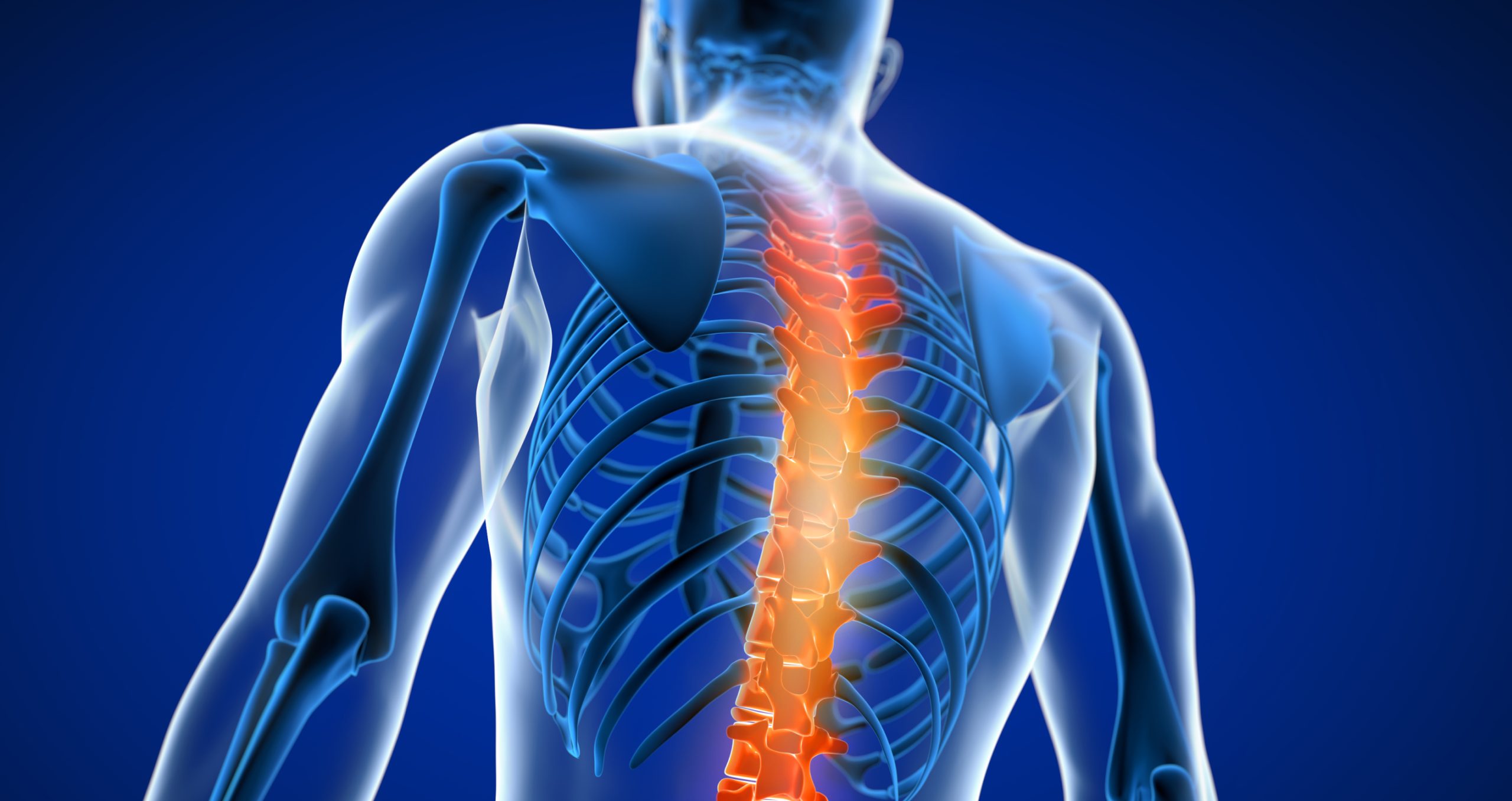
Accurately diagnosing soft tissue injuries within the neck and spine can be challenging. What’s more, subjective clinical assessments alone are not enough. According to a meta-analysis of 33 clinical studies, diagnostic imaging was not performed in patients presenting for care—contrary to guidelines—in 65.6% of patients with red flags and in 60.8% of patients with a clinical suspicion of lower back pathology.1
Most attorneys now know that MRI is the recommended initial imaging modality for diagnosing traumatic fracture and soft tissue injury due to its higher sensitivity. It is superior to other imaging modalities (like X-ray) for detecting anomalies of the spinal cord, bulging discs, small disc herniations, pinched nerves and a myriad of soft tissue problems.2 In fact, MRI has achieved reported sensitivities for intervertebral disc injury of 93%, posterior longitudinal ligament injury of 93% and interspinous ligament injury of 100%.3
“Using MRI as an initial diagnostic step for neck and back injuries can not only affirm an initial diagnosis, but may reveal additional pathology,” says Dr. Sana Khan, Medical Director of Expert MRI. “A quality MRI study provides the objective evidence for insurance companies that may be necessary to ensure the patient receives the full amount of care needed for their recovery.”
Khan notes, however, that not all MRI providers are adept at finding the full extent of injury. “Traditional MRI is powerful and accurate, however it fails more than 50% of the time when it comes to identifying cervical spine and ligament damage caused by a whiplash injury.”
Khan claims that the two key elements are needed to identify the true breadth of these injuries. “First, you need a team of radiologists experienced in this type of musculoskeletal diagnosis. Experience really does matter, especially as it relates to soft tissue injury. Second, you need the right imaging tools to identify the injury. In my experience, you need the option to image the patient in multiple positions that are not possible with supine MRI systems, which image the patient when they are lying down. If the pain is only present when the patient is standing, traditional MRI may never identify the problem.”
In addition to high-field MRI, Dr. Khan’s team uses upright MRI systems. These can often reveal problems that would otherwise not have been found.
“Having the right MRI tools at your disposal is so vitally important to diagnosing the totality of injury. When the full extent of the injury is documented, it becomes much easier for that patient to receive the full amount of insurance coverage needed for their treatment, which can often take many months of therapy. And for an accident/injury case, this ensures the attorney is able to maximize the amount of award for their client, to ensure they are treated properly.”
- Jenkins H, Downie AS, Maher CG, Moloney NA, Magnussen JS, Hancock MJ. Imaging for low back pain: is clinical use consistent with guidelines? A systematic review and meta-analysis [published online January 5, 2018]. Spine Journal. doi: 10.1016/j.spinee.2018.05.004
- Maher C, Underwood M, Buchbinder R. Non-specific low back pain. Lancet. 2017;389(10070):736–747. doi: 10.1016/S0140-6736(16)30970-9.
- Goradia D, Linnau KF, Cohen WA, Mirza S, Hallam DK, Blackmore CC. Correlation of MR imaging findings with intraoperative findings after cervical spine trauma. Am J Neuroradiol2007;28:209-15.


