CT—also known as a ‘Cat Scan’—stands for computed tomography. It is a highly useful innovation to see inside the body non-invasively. Most people are not aware that the lifesaving CT scanning technology actually has its roots in rock and roll.
In the 1960s, it became apparent to doctors that a more precise and detailed imaging technology was needed. Until then, X-rays were the primary way to see inside the body. While X-rays are fine for diagnosing certain health conditions, it was woefully inadequate when it came to seeing detail within the body’s internal organs, brain and soft tissue structures like cartilage.
Electric and Music Industries (EMI) is the company that owned the Abbey Road Studies in the 1960s and 1970s. The incredible popularity of the Beatles during that time produced record profits for the company, which they were able to reinvest to fund various R&D projects. In 1972—the year The Essential Beatles album was released—British engineer Godfrey Hounsfield of EMI Laboratories, working with physicist Dr. Allan Cormack, developed the first commercially available CT scanner from this funding. For their significant achievement, both were awarded the Nobel Prize in Physiology and Medicine in 1979.
The principle of CT scanning was as ingenious as it was elegant. Hounsfield, a high school dropout, said the idea came to him while he was on vacation. He was thinking about a way to reconstruct a 3D image of a box. He started re-imagining the box as a series of “slices” instead of the whole box.
At EMI, Hounsfield and Cormack refined the concept of using multiple X-rays to generate individual “slices” of human anatomy, which could then be reconstructed by a computer in three dimensions. While conventional X-ray uses a fixed tube that sends the X-rays in a single direction, this new approach would require sending focused X-rays from multiple angles around the body. The inventors accomplished this by fixing an X-ray camera to a rotating disc that could pass around the patient’s body, focused at a detector that is also rotating around the patient. That accounts for the scanner’s signature “donut” shape that remains today.
During a CT scan, the rotating camera captures hundreds of digital X-ray images forming the anatomical “slices” that a radiologist views on a special computer. The computer allows the radiologist to view the individual slices, or seamlessly “move” their view through the area imaged with 3D reconstruction, to look for abnormalities and form a diagnosis.
Over time, CT images have improved, as have 3D reconstruction capabilities. Modern CT scanners are faster, require the patient to remain in the scanner for only a short period of time and expose patients to less radiation. Today, CT images are used to diagnose brain tumors, problems with the bones, joints and muscles, heart disease and coronary blockages, internal bleeding, and cancer within nearly every organ system in the body. The 3-dimensional reconstruction capabilities of CT assists surgeons in pre-surgical planning. It is also used to guide biopsies and various forms of disease treatment from inside the body.
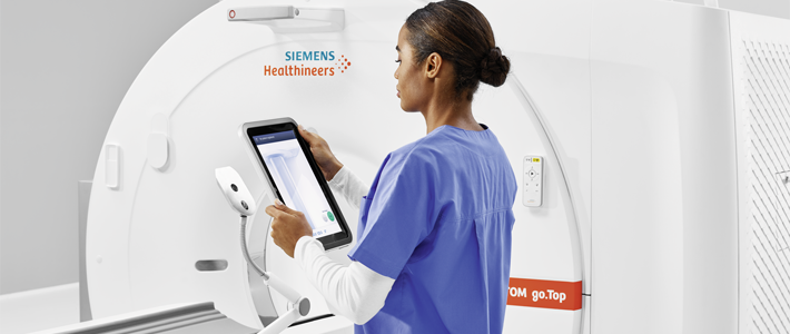
With a name like ExpertMRI, it isn’t obvious that we use CT technology… but we do. We operate some of the most advanced CT scanners available today at several of our facilities. They are an excellent complement to technologies like MRI and ultrasound and have their place in diagnosing injuries to the brain and musculoskeletal system.