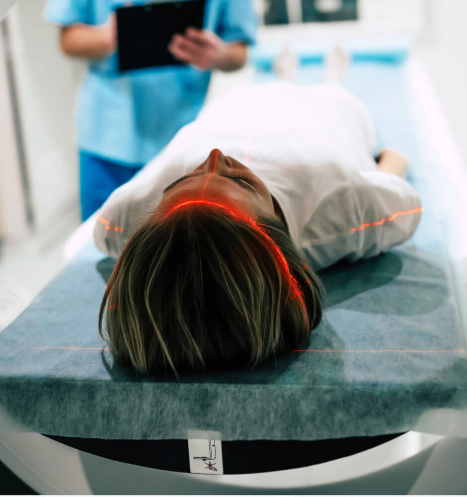
• Skip to main content

Specialized Services

  • High-Field Strength MRI
  • Front open mri
  • Computed Tomography
  • X-Ray
Find Nearest Expert MRI Location

Coronavirus icon for Expert MRICorona Virus Update Go Here

  • Patients
  • Physicians
  • Attorneys
  • About Us
  • Blog
  • Facebook profile and Facebook icon for Expert MRI
  • YouTube profile and YouTube icon for Expert MRI
  • Instagram profile and Instagram icon for Expert MRI
  • LinkedIn profile and LinkedIn icon for Expert MRI
  • Insurance
  • Services
    • High-Field Strength MRI
    • Front open mri
    • X-Ray
    • Computed Tomography
    • Ultrasound
    • Interventional Radiology
  • Locations
  • Access Portal
  • Patients
  • Physicians
  • Attorneys
  • About Us
  • Blog
  • Facebook profile and Facebook icon for Expert MRI
  • Instagram profile and Instagram icon for Expert MRI
  • YouTube profile and YouTube icon for Expert MRI
  • LinkedIn profile and LinkedIn icon for Expert MRI

Technology | Science | Evidence

  • Insurance
  • Services
    • High-Field Strength MRI
    • Front open mri
    • X-Ray
    • Computed Tomography
    • Ultrasound
    • Interventional Radiology
  • Locations
  • Access Portal

Uncategorized

False Negative MRIs: What are they and how to avoid them?

Simply put, a “false negative” MRI is an MRI exam that fails to reveal a problem that is most certainly there. It’s far more common than most people think.

According to the American Medical Association, “In up to 85% of individuals who report back pain, no pain-producing pathology can be identified…” For an individual experiencing back pain, this can be incredibly frustrating. To them, the pain is real, and without evidence, getting the right care—and getting the coverage for that care—can be a challenge.

Why does this happen? With today’s modern MRI machines, finding the source of pain seems like a foregone conclusion. But in reality, the inability to identify pain pathology is a failure on several fronts.

Misinterpreted Images

MRI exams—as well as all radiology exams—can be misinterpreted by the radiologist for a variety of reasons. A false negative diagnosis can lead the referring doctor and their patient down the incorrect path and delay critical treatment. If you’ve experienced an injury to the neck or spine, it’s important to have a radiologist experienced with these types of injuries. Their specialization means they are more likely to find problems that could be missed by a radiologist who, for example, mostly reads mammograms.

Scanning in the wrong position

Some injuries to the spine cause the patient great pain when sitting, standing or bending over, but not necessarily while lying flat on their back. Unfortunately, most MRI machines are designed to image the patient in the recumbent (lying down) position. Only upright MRI systems are capable of imaging the patient in multiple positions, including those positions in which they are experiencing the most pain.

Scanning in the wrong location

connect the skull to the cervical spinal column. These are considered “major stabilizers” of the head and neck. Many MRIs of the spine do not include the craniocervical junction that contains these ligaments, and can therefore miss an injury or the full extent of the injury. It is not uncommon for a patient to receive a normal MRI yet still be experiencing dizziness or “drop attacks” because of a missed injury within the craniocervical junction.

Ligamentous injury measures are not done

When it comes to soft tissue injury, such as damage to the ligaments and tendons in the shoulder or knee, important clinical insights can be gained when these soft tissue strain patterns are properly measured. This can lead to a better understanding of the loss of motion resulting from the injury, which can be used to plan more effective rehabilitation and treatment. MRI is capable of making these in vivo strain measurements, but not all imaging centers take the time to perform them.

Scanning at the wrong time

Scans performed within 1 month of a musculoskeletal injury can be more accurate than those performed between 1 and 2 months. This is because scar tissue develops, which can lead to a discrepancy in the type of injury being diagnosed. In addition, performing MRI imaging immediately after a suspected traumatic brain injury (TBI) can result in more accurate and better detection of microbleeds on the brain or cerebral microhemorrhages.

Using the wrong MRI sequences

Conventional MRI sequences may not demonstrate certain imaging changes well. In the example of head trauma, specialized sequences may be required. These include susceptibility-weighted imaging (SWI) for evaluating smaller hemorrhages within the brain, diffusion-weighted imaging (DWI) for evaluating ischemic stroke and early infarcts, and diffusion tensor imaging (DTI) for the assessment of diffuse axonal injury.

Injuries of significance are unrecognized by the referring doctor

If a doctor refers you to an MRI for suspected disc herniation, and that finding is confirmed on MRI, there is sometimes a tendency to stop reading right there… and not look at other significant findings within the exam report. Atypical disc herniations can also lead to other conditions, such as a cerebrospinal fluid leak, acute calcific discitis, cysts within the discs and more, which may require different or additional treatment strategies.

If you are a patient, having your MRI at Expert MRI greatly reduces the chance for a false negative— which in turn can reduce frustration, eliminate the need for additional testing and save you valuable time. If you are a physician or attorney and would like more detail on our approach to reducing false negatives, please contact us to arrange a presentation.

Pediatric Scoliosis Testing – A “Real Game-Changer”

recently, the diagnosis of scoliosis in children and adolescents required a series of X-rays of the full spine to measure the Cobb angle—the degree of curvature of the spine that could indicate a disorder. To monitor scoliosis progression or the effectiveness of treatment, these patients may also require up to 4-5 additional X-rays per year for a period of 3-5 years before the child reaches skeletal maturity.

Unfortunately, the use of repeated and numerous scoliosis X-rays on children have been linked to higher incidences of breast cancer, thyroid cancer and leukemia.1,2  Even today’s lower-dose radiographic techniques cannot eliminate long-term risk.

“It is widely known that young patients have higher radiosensitivity than adults, meaning that their organs are susceptible to damage from radiation and a greater risk of cancer later in life,” says Dr. Andrew Thierry, a musculoskeletal radiology specialist at Expert MRI. “It was clear to us that a safer test was needed during this critical point in their development.

The challenge for Dr. Thierry and his colleagues at Expert MRI was to find a way to use MRI—a superior imaging technique for spine imaging that doesn’t use iodizing radiation—for scoliosis testing. The problem is that most MRI systems image the patient while he or she is lying on their back, and scoliosis imaging requires the patient to be sitting or standing in an upright, weight-bearing position.

Their solution was to use upright MRI, an “open” MRI technology that is ideal for pediatric imaging because it does not require the child to be imaged in a small, confined tube-like space. The Expert MRI team began scanning children several years ago and have since refined the imaging technique and protocols into a practical, well-tolerated and inexpensive seven-minute test to accurately measure spine deformity.

“This is a real game changer, and I don’t use that term lightly,” adds Dr. Thierry. “In addition to being safer for kids, the MRI test is more precise and enables us to see the soft tissues surrounding the spine. This means we can evaluate related conditions such as muscle atrophy and the impact of the scoliosis on internal organs.”

Expert MRI is the only imaging center network in California to provide this innovative and safe scoliosis test. Doctors and parents are encouraged to specify this test instead of x-rays for children in need of both initial evaluation and ongoing progression monitoring of scoliosis.

“The kids don’t mind having this test at all,” Dr. Thierry adds. They get to sit and watch cartoons while they are in the scanner. And if the patient is very young, the MRI technology is so safe that a parent can even sit inside the machine with their child during the test.”

For more information about Expert MRI’s scoliosis imaging, please call (877) 674-8888.

1. Doody M, Lonstein JE, Stovall M, Hacker DG, Luckyanov N, Land CE. Breast cancer mortality after diagnostic radiography: findings from the U.S. Scoliosis Cohort Study. Spine 25:2052-63, 2000. 2. Rao, PS, Gregg EC. A revised estimate of the risk of carcinogenesis from x-rays to scoliosis patients. Invest Radiol 19:58-60, 1984.

MRI vs CT: Which type of scan is right for my condition?

CT (computed tomography) and MRI (magnetic resonance imaging) are imaging tests used to produce internal images of the bones, organs, structures, and tissues within the body. The machines look similar, resembling large donuts into which the patient is placed during the exam. To the untrained eye, even the images they generate appear similar.

In reality, these are two very different technologies and are mostly used in different ways by healthcare professionals to reach an accurate diagnosis for a patient. Because the technology of each continues to evolve, it is not uncommon for one of these technologies to replace the other as a doctor’s preferred imaging technique.

CT scans are used more frequently than MRIs. They are also generally less expensive. A CT scanner is basically an X-ray camera rotating around the patient, capturing multiple images of a part of the body from different angles. These images are then assembled by a computer into anatomical “slices” that are reviewed by a radiologist—a specially trained medical doctor—who will provide a diagnosis. CT is fast, requiring only a few seconds to capture the images. Because the scan is X-ray-based, it exposes the patient to a low amount of radiation.

MRI on the other hand, uses a powerful magnet, radio waves, and a sophisticated computer to produce its images. In many cases, MRI images are more detailed, especially when it comes to soft tissues within the body like tendons and ligaments. However, an MRI takes much longer than a CT scan and is typically more expensive. Because of this, CT is often used first to diagnose or rule out certain conditions, such as cancer. It is also more often used for cardiac imaging, where the CT scanner’s incredibly fast speed is able to capture images of the heart and arteries between heartbeats.

While X-ray is still used for broken bones, it is not recommended for complex injuries of the brain, neck or spine. MRI is the preferred method for these types of injuries, where diagnosing the full extent of injuries to the soft tissues and structures surrounding the bones is just as important as diagnosing injuries to the bones themselves. This is why MRI is used most often for those who have experienced an injury; a CT scan will not reveal soft tissue injuries common to whiplash, traumatic brain injury, and more.

Furthermore, while the radiation exposure of a single CT scan is low, many in the medical field have concluded that repeated CT scanning—especially in children—could be harmful. This is why MRI is preferred for children (e.g. scoliosis imaging) and women who are pregnant (e.g. breast screening). Conversely, MRI cannot be used with some patients who have certain types of implanted medical devices that contain metal, such as pacemakers and brain stimulators. Fortunately, newer implant technologies are designed with MRI imaging in mind, so patients with these devices may be safely imaged within a magnetic field.

In most cases, your doctor will know what test to order for your specific needs. When the decision is not certain, your doctor may consult with one of the doctors at Expert MRI to recommend the best type of scan for your condition or suspected condition. 

Expert MRI has California’s widest assortment of high field, open and upright MRI systems to give clinicians more imaging options to achieve the most accurate diagnosis for their patients. In addition, the cash price of our MRI exams is very low, in many cases lower than the cost of a CT scan at a hospital or other imaging center.

Expert MRI
  • Facebook profile and Facebook icon for Expert MRI
  • Instagram profile and Instagram icon for Expert MRI
  • YouTube profile and YouTube icon for Expert MRI
  • LinkedIn profile and LinkedIn icon for Expert MRI

Services

  • High-Field Strength MRI
  • Front open mri
  • Computed Tomography
  • X-Ray
  • Ultrasound
  • Interventional Radiology

Locations

  • Bellflower
  • Bakersfield
  • San Gabriel
  • Sherman Oaks
  • Winnetka
  • Riverside
  • Beverly Hills
  • Culver City
  • Fullerton
  • Costa Mesa

Company

  • About Us
  • Services
  • Blog
  • Physician
  • Patients
  • Insurance
  • Attorney
  • Careers

Legal

  • Privacy policy
  • Disclaimer
  • HIPAA
  • Anti-Discriminatory Policy

© All rights reserved | 2016 - 22 | Powered by Quadrant Technology

Schedule An Appointment

"*" indicates required fields

MM slash DD slash YYYY