In recent years, the field of medical imaging has made significant strides, offering healthcare professionals powerful tools to diagnose and manage various conditions with greater precision. One such advanced technique is Diffusion Tensor Imaging (DTI), a specialized form of MRI that maps the pathways of the nervous system. DTI has revolutionized our understanding of the brain’s intricate wiring and its applications in diagnosing neurological disorders. In this article, we will explore the science behind DTI, its applications, and how it benefits both patients with neurological conditions and the healthcare professionals who care for them.
Understanding Diffusion Tensor Imaging (DTI)
Diffusion Tensor Imaging (DTI) is an MRI-based technique that measures the diffusion of water molecules in the brain. Water molecules naturally move in random directions, a process known as Brownian motion. In the brain, however, the movement of water molecules is not entirely random; it is influenced by the structures through which they pass. In white matter, the brain’s communication network, water molecules tend to diffuse along the direction of nerve fibers rather than across them. This directional diffusion is known as anisotropy, and it is the basis for DTI.
DTI provides a way to visualize and measure the orientation and integrity of white matter tracts in the brain. By applying mathematical models to the diffusion data, DTI generates images that depict the pathways of these tracts, often referred to as “tractography.” These images can reveal crucial information about the brain’s connectivity, which is invaluable in both research and clinical settings.
The Importance of White Matter in Neurological Health
White matter consists of bundles of axons, the long projections of nerve cells (neurons) that transmit electrical signals between different regions of the brain and between the brain and the spinal cord. The integrity of white matter is essential for efficient communication within the nervous system. Damage to white matter can disrupt this communication, leading to a variety of neurological symptoms.
Conditions such as multiple sclerosis (MS), traumatic brain injury (TBI), stroke, and neurodegenerative diseases like Alzheimer’s and Parkinson’s disease can affect white matter. DTI is particularly valuable in assessing the extent of white matter damage in these conditions, providing insights that traditional MRI techniques might miss.
How DTI Works: The Science Behind the Imaging
The process of DTI involves several steps. First, the MRI scanner uses magnetic fields and radio waves to create detailed images of the brain. During the scan, the movement of water molecules is tracked in multiple directions, capturing data about how they diffuse through the brain’s tissues. This diffusion data is then used to calculate two key metrics:
- Fractional Anisotropy (FA): This metric quantifies the degree of anisotropy or directional dependence of water diffusion. Higher FA values indicate more organized and intact white matter tracts, while lower FA values suggest damage or disruption.
- Mean Diffusivity (MD): MD measures the overall movement of water molecules in the brain, providing information about tissue density and cellular structure.
These metrics are combined to generate tractography images, which visually represent the brain’s white matter pathways. The resulting images are not only visually striking but also clinically informative, offering a non-invasive way to assess brain health.
Applications of DTI in Neurology
DTI has a wide range of applications in the diagnosis and management of neurological conditions. Here are some of the key areas where DTI has proven particularly valuable:
- Multiple Sclerosis (MS)
Multiple sclerosis is a chronic autoimmune disease that affects the central nervous system, leading to the destruction of myelin, the protective sheath around nerve fibers. This myelin damage disrupts communication between the brain and the rest of the body, resulting in a wide range of symptoms.
DTI can detect early changes in white matter integrity that may not be visible on conventional MRI scans. By assessing the FA values in different regions of the brain, neurologists can monitor the progression of MS and evaluate the effectiveness of treatments. DTI has also been used to study the relationship between white matter damage and cognitive impairment in MS patients.
- Traumatic Brain Injury (TBI)
Traumatic brain injury is a leading cause of disability worldwide, often resulting from accidents, falls, or sports-related injuries. Even mild TBI, commonly known as a concussion, can cause subtle changes in brain structure that are difficult to detect with standard imaging techniques.
DTI is particularly sensitive to diffuse axonal injury, a type of brain damage that occurs when the brain’s white matter is stretched or sheared. This type of injury is common in TBI and can lead to long-term cognitive and emotional difficulties. By identifying areas of reduced FA, DTI helps clinicians assess the severity of TBI and guide rehabilitation strategies.
- Stroke
A stroke occurs when blood flow to a part of the brain is interrupted, leading to brain tissue damage. The extent and location of the damage determine the symptoms and the patient’s recovery prospects.
DTI can provide valuable information about the integrity of white matter tracts following a stroke.
This information can help predict a patient’s recovery potential and guide rehabilitation efforts. For example, DTI can identify which motor pathways are still intact, aiding in the development of personalized physical therapy programs.
- Neurodegenerative Diseases
Neurodegenerative diseases, such as Alzheimer’s and Parkinson’s disease, are characterized by the progressive loss of neurons and their connections. DTI has been used to study the changes in white matter that occur in these conditions, offering insights into disease mechanisms and progression.
In Alzheimer’s disease, for instance, DTI has revealed disruptions in the white matter tracts that connect memory-related regions of the brain. This information can complement other diagnostic tools, such as PET scans and cognitive testing, to provide a more comprehensive assessment of the disease.
- Surgical Planning and Navigation
In neurosurgery, precise knowledge of the brain’s anatomy is crucial to avoid damaging critical areas during surgery. DTI can map out the white matter tracts surrounding a tumor or lesion, allowing surgeons to plan their approach more effectively. By preserving key pathways, DTI-guided surgery can help reduce postoperative complications and improve patient outcomes.
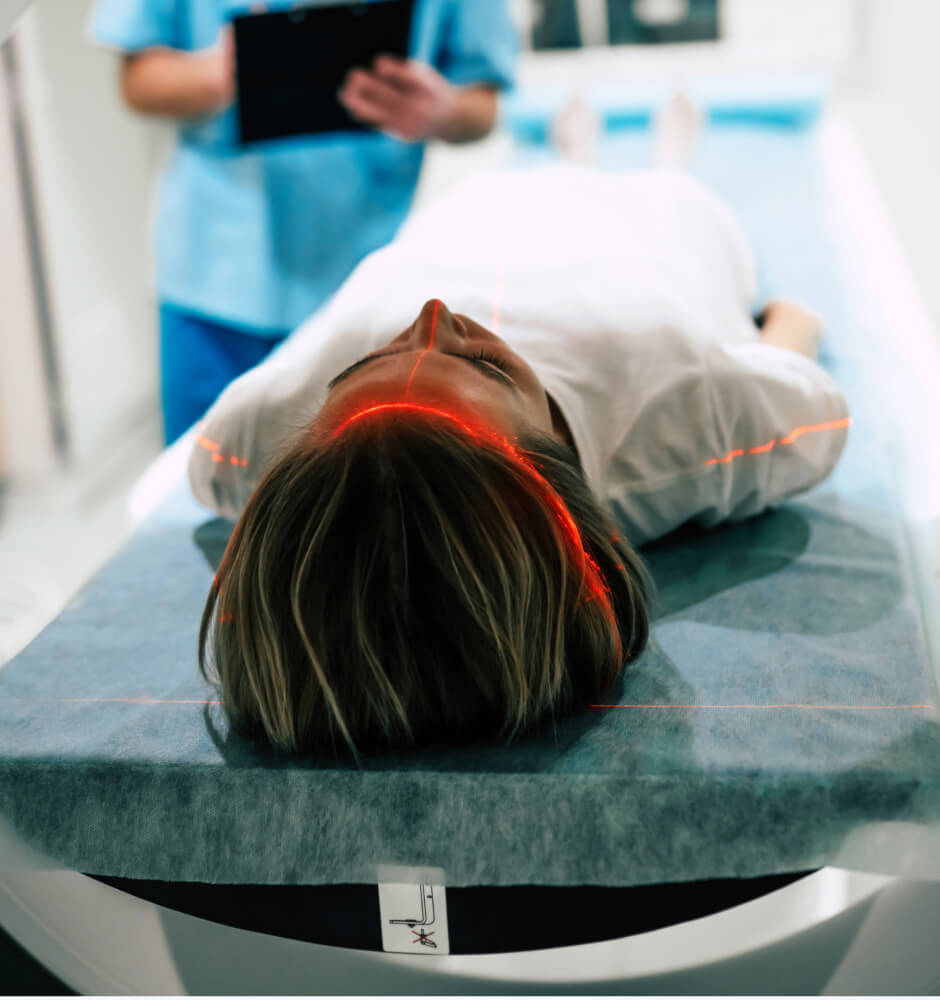
The Patient’s Perspective: What to Expect from a DTI Scan
For patients undergoing a DTI scan, the experience is similar to that of a standard MRI. The patient lies still on a table that slides into the MRI machine, which takes a series of images over 30 to 60 minutes. The procedure is non-invasive and does not involve radiation, making it a safe option for most patients.
The primary difference between a DTI scan and a conventional MRI is the type of information obtained. While a standard MRI provides detailed images of the brain’s structure, DTI adds another layer of information by revealing how water molecules move through the brain’s tissues. This additional data can be crucial in diagnosing and managing neurological conditions.
Patients with neurological disorders often experience uncertainty and anxiety about their condition. The ability of DTI to provide detailed insights into the brain’s connectivity can offer reassurance by enabling more accurate diagnoses and more personalized treatment plans. For example, a patient with MS may benefit from regular DTI scans to monitor disease progression and adjust therapy accordingly.
The Healthcare Professional’s Perspective: Integrating DTI into Clinical Practice
For neurologists and other healthcare professionals, DTI offers a powerful tool to enhance diagnostic accuracy and patient care. By providing detailed information about the brain’s white matter, DTI can uncover abnormalities that may not be visible on conventional MRI scans. This capability is particularly valuable in complex cases where the diagnosis is uncertain.
Integrating DTI into clinical practice requires collaboration between radiologists, neurologists, and other specialists. Radiologists play a key role in interpreting DTI results and generating tractography images, while neurologists use this information to make informed decisions about diagnosis and treatment. In some cases, DTI findings may prompt further testing or a referral to a specialist.
For healthcare professionals, staying informed about the latest advances in medical imaging is essential. DTI is a rapidly evolving field, with ongoing research exploring new applications and refining existing techniques. By incorporating DTI into their practice, healthcare providers can offer patients access to cutting-edge diagnostics and improve overall care quality.
The Future of DTI: Advancing Neurological Diagnostics
As research continues, the applications of DTI are likely to expand further, offering new insights into the brain’s structure and function. For instance, ongoing studies are investigating the use of DTI in psychiatric disorders such as schizophrenia and bipolar disorder, where white matter abnormalities may play a role.
In addition to clinical applications, DTI is also being used in research to explore fundamental questions about brain development, aging, and plasticity. By tracing the pathways of the nervous system, DTI is helping to unravel the complexities of the brain and improve our understanding of neurological health.
Diffusion Tensor Imaging (DTI) represents a significant advancement in the field of neurological diagnostics. By providing detailed images of the brain’s white matter pathways, DTI offers valuable insights into a wide range of conditions, from multiple sclerosis and traumatic brain injury to stroke and neurodegenerative diseases. For patients with neurological disorders, DTI can lead to more accurate diagnoses and more personalized treatment plans, offering hope and reassurance in the face of uncertainty.
For healthcare professionals, DTI is a powerful tool that enhances diagnostic accuracy and improves patient care. By staying informed about the latest advances in DTI and incorporating it into clinical practice, healthcare providers can offer patients access to cutting-edge diagnostics and contribute to the ongoing advancement of neurological medicine.
As DTI technology continues to evolve, its potential applications will likely expand, offering new opportunities to improve our understanding of the brain and enhance patient outcomes. For now, DTI stands as a testament to the power of medical imaging to transform the way we diagnose and treat neurological conditions, paving the way for a healthier future.