Table of Contents
Unless you have had a CT scan before, you might not know exactly what it is. Even if you’ve had a CT scan, you might not know how it actually works. Today, we’ll explain everything with the help of two carbohydrate-laden foods: Donuts and sliced bread.
First, it’s important to understand that a CT scanner is based on x-ray technology. With a conventional x-ray, the patient lies on a table. A single, narrow x-ray beam is released from a special tube that hangs above the part of the patient being imaged. Beneath the patient is a special detector that collects the image. In the past, there was film in the detector that collected the image, just like with an old-style photograph. With today’s digital x-rays, the image is collected on a digital detector and is then transferred to a computer which the radiologist—a specially trained doctor—uses to diagnose health conditions like broken bones.
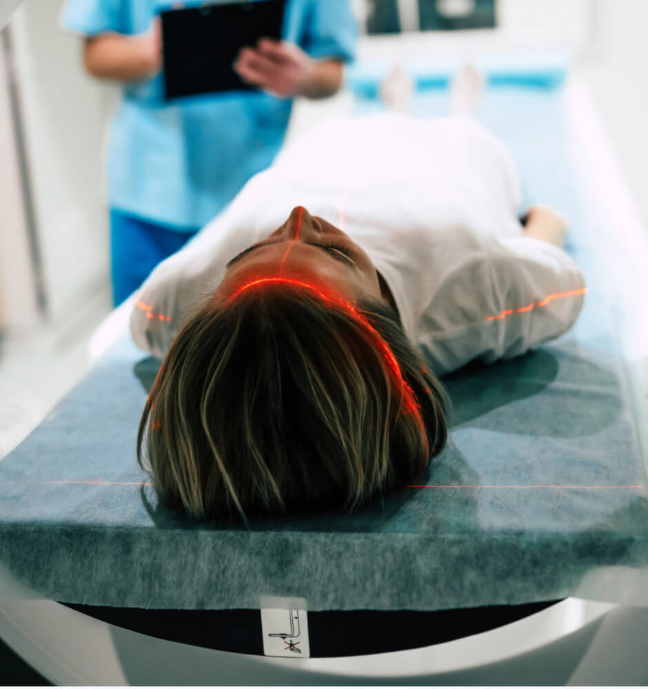
But in a CT scanner, the x-ray tube and the detector are both inside the “donut” shaped housing that the patient is guided into for the examination. During a CT scan, a motorized x-ray tube revolves completely around the patient again and again, sending out multiple x-rays while it rotates. On the opposite side of the tube, also revolving around the patient, is the digital x-ray detector. Many images are obtained, and each individual x-ray image is collected, organized and stored in a computer.
Now, here’s where the bread reference comes in. Each x-ray image represents a very thin “slice” of the patient’s anatomy. When these images are combined together by the computer, an even more accurate representation of the part of the patient’s body being imaged becomes clear. Instead of just viewing a single slice, the doctor can see the entire loaf, so to speak.
The benefits of multi-slice CT imaging
By being able to look at anatomy at various depths, the radiologist gets a more complete view of the inside of the body, which is why CT scans are used to diagnose disease in the body’s organs, identify various orthopedic conditions, evaluate the brain for injury and more. The images can also be reconstructed in three dimensions for a more complete understanding of the nature of the disease or injury, as well as to assist in surgical planning, if necessary.
In some cases, a patient may be injected with a substance called a contrast agent before their CT scan. Contrast is used to differentiate between certain organs and improve the detection or characterization of certain diseases. It is also used to evaluate blood flow through the body’s blood vessels. For example, a CT scan of the heart with contrast can help to evaluate blood flow through the coronary arteries and help determine if there is a blockage that is putting the patient at risk for a heart attack. This type of CT scan may be called CT perfusion imaging or CT angiography (CTA).
CT scans are also now widely used for preventive imaging. Unlike diagnostic CT scans, preventive imaging evaluates otherwise healthy individuals. Just like a mammogram is used to find cancer at an earlier, more treatable stage, CT scans can be used to evaluate the body’s organs, such as the heart, lungs, kidneys, brain and other organs to find disease early, giving people the chance to treat it before symptoms appear and it becomes life-threatening.
The more slices, the better
Since the 1970s, CT scanners have constantly evolved to attain better image quality. Better image quality is achieved by producing smaller and more numerous image “slices,” which is why CT scanner models are named for the number of image slices. For example, an 8-slice CT captures eight slices of data with each rotation of the scanner around the patient. The modern 64-slice CT scanners used by Expert MRI generate 64 slices of data with each rotation, meaning that more numerous, thinner images are captured faster, which translates into greater image detail, faster scan times and a more comfortable experience for the patient.
Unlike other diagnostic imaging equipment, our 64-slice CT scanners feature an open design, which is not confining or claustrophobic for the patient. The image acquisition speeds are so fast that the scanner can accurately image the heart between individual beats.
Do the additional image slices generated by CT mean more radiation exposure?
Yes, CT scans expose patients to more radiation than an x-ray. However, the amount of radiation received is still very small and within acceptable safety limits for humans. In addition, each new generation of CT equipment is faster, which means less overall radiation exposure for the patient. Finally, the radiologists and technologists at Expert MRI have been trained in the use of special protocols designed to obtain optimized images at the lowest possible radiation dose. In short, the diagnostic value of the images provided by CT far outweighs the potential risks of radiation exposure.
Is it a CT scan or a CAT scan?
Finally, you may be wondering, “Why is it called a CT scan, or in some cases, a “CAT” scan?
CT stands for computed tomography. The word “tomography” comes from the Greek word “tomos” which means section or slice and “graphe” which means drawing. When the technology was first developed, its name was computed axial tomography, which is why some people may still refer to the imaging test as a CAT scan. However, it has since been shortened to CT for simplicity.
If you’ve read this far, you now know more about CT scans than most people! If you’re curious, we have a short, animated video on our website that demonstrates how a CT scanner works. You can view the video here.


