

Table of Contents
Simply put, a false negative MRI is an MRI exam that fails to reveal a problem that is most certainly there. It’s far more common than most people think.
According to the American Medical Association, “In up to 85% of individuals who report back pain, no pain-producing pathology can be identified…” For an individual experiencing back pain, this can be incredibly frustrating. To them, the pain is real, and without evidence, getting the right care—and getting the coverage for that care—can be a challenge.
Why does this happen? With today’s modern MRI machines, finding the source of pain seems like a foregone conclusion. But in reality, the inability to identify pain pathology is a failure on several fronts.
Misinterpreted Images
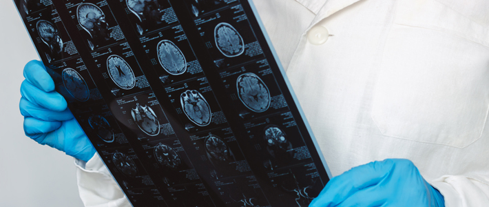
MRI exams—as well as all radiology exams—can be misinterpreted by the radiologist for a variety of reasons. A false negative diagnosis can lead the referring doctor and their patient down the incorrect path and delay critical treatment. If you’ve experienced an injury to the neck or spine, it’s important to have a radiologist experienced with these types of injuries. Their specialization means they are more likely to find problems that could be missed by a radiologist who, for example, mostly reads mammograms.
Scanning in the wrong position
Some injuries to the spine cause the patient great pain when sitting, standing or bending over, but not necessarily while lying flat on their back. Unfortunately, most MRI machines are designed to image the patient in the recumbent (lying down) position. Only upright MRI systems are capable of imaging the patient in multiple positions, including those positions in which they are experiencing the most pain.
Scanning in the wrong location
connect the skull to the cervical spinal column. These are considered “major stabilizers” of the head and neck. Many MRIs of the spine do not include the craniocervical junction that contains these ligaments, and can therefore miss an injury or the full extent of the injury. It is not uncommon for a patient to receive a normal MRI yet still be experiencing dizziness or “drop attacks” because of a missed injury within the craniocervical junction.
Ligamentous injury measures are not done
When it comes to soft tissue injury, such as damage to the ligaments and tendons in the shoulder or knee, important clinical insights can be gained when these soft tissue strain patterns are properly measured. This can lead to a better understanding of the loss of motion resulting from the injury, which can be used to plan more effective rehabilitation and treatment. MRI is capable of making these in vivo strain measurements, but not all imaging centers take the time to perform them.
Scanning at the wrong time
Scans performed within 1 month of a musculoskeletal injury can be more accurate than those performed between 1 and 2 months. This is because scar tissue develops, which can lead to a discrepancy in the type of injury being diagnosed. In addition, performing MRI imaging immediately after a suspected traumatic brain injury (TBI) can result in more accurate and better detection of microbleeds on the brain or cerebral microhemorrhages.
Using the wrong MRI sequences
Conventional MRI sequences may not demonstrate certain imaging changes well. In the example of head trauma, specialized sequences may be required. These include susceptibility-weighted imaging (SWI) for evaluating smaller hemorrhages within the brain, diffusion-weighted imaging (DWI) for evaluating ischemic stroke and early infarcts, and diffusion tensor imaging (DTI) for the assessment of diffuse axonal injury.
Injuries of significance are unrecognized by the referring doctor
If a doctor refers you to an MRI for suspected disc herniation, and that finding is confirmed on MRI, there is sometimes a tendency to stop reading right there… and not look at other significant findings within the exam report. Atypical disc herniations can also lead to other conditions, such as a cerebrospinal fluid leak, acute calcific discitis, cysts within the discs and more, which may require different or additional treatment strategies.
If you are a patient, having your MRI at Expert MRI greatly reduces the chance for a false negative— which in turn can reduce frustration, eliminate the need for additional testing and save you valuable time. If you are a physician or attorney and would like more detail on our approach to reducing false negatives, please contact us to arrange a presentation.